What is retrochorial hematoma? Retrochorial hematoma - causes and symptoms of the disease, diagnosis and manifestations, treatment methods
Retrochorial hematoma during pregnancy is an accumulation of blood in the uterus that occurs as a result of partial rejection of the fertilized egg in the early stages of gestation. This condition threatens the normal development of the embryo and can result in miscarriage.
Timely medical care makes it possible to maintain a pregnancy. Every expectant mother needs to know the first signs of this pathology in order to avoid serious consequences.
The embryo is surrounded by the chorionic membrane. During the intrauterine development of the fetus, it participates in the formation of the placenta. If, due to the influence of any negative factors, chorion begins to be rejected from the uterine mucosa, bleeding may begin. In this case, blood gradually accumulates behind the chorionic membrane in the resulting cavity between it and the inner surface of the uterus.
The diagnosis of “retrochorial hematoma” can only be made before the 16th week of pregnancy. Later, placental tissue should already be formed, so in later stages a similar process is called placental abruption.
Regardless of the stage of pregnancy, these phenomena are very dangerous for both the woman and the fetus. Often the disease is asymptomatic, which is why it is so important to visit a gynecologist regularly.
Causes
There are many reasons for this pathology. These include:
- hormonal disorders;
- mechanical damage (bruises, abdominal injuries);
- severe stress;
- severe early toxicosis;
- alcohol abuse, smoking;
- malformations of the reproductive organs;
- diseases of the uterus (, polyps,);
- exacerbation of chronic diseases of the genitourinary system;
- blood diseases;
- cardiovascular diseases;
- autoimmune conditions;
- endocrine diseases;
- abnormalities of the developing embryo;
- working with hazardous substances;
- physical overload, especially heavy lifting.
Symptoms
In the early stages, the following complaints arise: aching pain in the lower abdomen with intermittent irradiation to the lower back, vaginal discharge of various types. The appearance of the first signs of pathology is a reason for urgent medical consultation.
At the beginning of the disease, a woman is bothered by scarlet bloody discharge from the genital tract. They indicate the beginning of detachment of the fertilized egg from the uterine mucosa. Dark, brownish spotting from the vagina can be an encouraging sign that bleeding has stopped and the process of resolving blood clots has begun.

If the process of detachment of the ovum continues, the nature of the pain syndrome changes. The pain becomes severe, cramping, and the discharge remains bloody.
The woman’s general well-being changes: severe weakness appears, accompanied by fear and anxiety, blood pressure drops, and the heartbeat quickens. This condition is a medical emergency.
Diagnostics
To preserve the health of the woman and the unborn child, the diagnosis must be made as quickly as possible. A medical appointment is required, including a thorough history taking, listening to complaints and a gynecological examination of the patient.
The following laboratory tests are carried out:
- biochemical blood test (increased blood clotting due to increased concentration) should be monitored over time;
- hormonal profile (deficiency);
- general blood and urine tests;
- vaginal smear to clarify the microflora and exclude sexually transmitted diseases.
Ultrasound is performed vaginally and transabdominally. It is an indispensable method for detecting bleeding in the initial stages and diagnosing asymptomatic hematomas in the upper parts of the uterus.
Ultrasound data will help resolve the issue of emergency hospitalization. In addition to the hematoma, the doctor performing the ultrasound may see thickening of the uterine wall, which causes the fertilized egg to become deformed.
Treatment
Inpatient treatment is carried out, as there is a threat of miscarriage and uterine bleeding, so hospitalization cannot be refused. It is mandatory under the following circumstances: the release of fresh blood from the vagina, a violation of the general condition, severe pain.
In addition, hospitalization is indicated in the presence of concomitant somatic, endocrine or inflammatory diseases. Treatment also takes place in a hospital if the volume of blood accumulated in the uterine cavity is 20 ml or more.
The goals of treatment are to stop bleeding and resolve the resulting hematoma. The joint efforts of the woman and the attending doctor, their complete understanding and trust are important. Therapy with the selection of medications, determining their dose, frequency and duration of administration is prescribed by the attending physician. Self-medication can have irreparable consequences.
Medicines are prescribed in tablet form or administered parenterally, which is determined by the severity of the woman’s condition. Local intravaginal use of drugs in the form of suppositories is also important.
The following groups of medicinal substances are used:
- analgesics;
- antispasmodics to reduce increased uterine tone;
- vascular drugs to improve blood circulation;
- hemostatics to stop bleeding;
- hormonal drugs;
- vitamins (ascorbic acid, vitamins A, E and group B);
- trace elements (potassium, magnesium, silicon, copper, calcium);
- sedative herbal;
- preparations containing iron.

Changing lifestyle and diet
If there is no pain or red bloody discharge, and the hematoma is small according to ultrasound, the observing gynecologist may allow the expectant mother to stay at home. But a woman must adhere to the recommended regimen, regularly visit the doctor and strictly follow all his instructions. Relatives need to take care of all household chores.
The expectant mother should observe bed rest, place a cushion under her lower back, creating an elevated position for the pelvis so that there is no stagnation of blood. It is necessary to eliminate worries and emotional stress, give up sexual activity, and normalize nutrition.
It is not recommended to consume foods that thin the blood - legumes, cabbage, coffee, carbonated drinks. It is worth giving preference to dishes rich in proteins and vitamins. It is also important to regulate your bowel movements and drink enough fluids.
Consequences of the disease
If a woman follows the doctor’s recommendations, she has every chance of recovering and keeping the child. Blood spilled beyond the chorionic membrane, with normalization of lifestyle and adequate therapy, gradually resolves within 15-35 days.
An embryo develops in the uterus in early pregnancy. It is protected by a thin film of the fertilized egg - the chorion, which during normal development is transformed into the placenta.
If during this period there is a failure in the development of the embryo, the embryonic egg is separated from its chorion film. The detachment site fills with blood. This pathology is called retrochorial hematoma during pregnancy. It can cause fetal development to stop and be lost.

Ultrasound can detect hematoma during pregnancy. In the photo taken by an ultrasound diagnostic device, the characteristic darkening is clearly visible, as well as the deformation of the fertilized egg.
A retrochorial hematoma develops during a singleton pregnancy at 5-7 weeks and in cases where a woman is pregnant with twins. Pathology can be suspected by the appearance of nagging pain in the lower abdomen, brownish or scarlet discharge. If a woman discovers these symptoms, she should immediately consult a doctor.
In addition to retrochorionic pathology, there are:
- subchorionic - it poses a serious threat to the fetus, surrounding the placenta;
- retroamniotic— timely detected formation, subject to adequate therapy, does not pose a threat to the developing baby;
- retroplacental- it is formed after the formation of the placenta, after 16 weeks of pregnancy;
- subamniotic— its presence is not dangerous for the baby, but requires regular monitoring;
- intrauterine— the presence of such a hematoma carries a serious risk for the fetus and requires urgent treatment.
Danger for the expectant mother and baby
If a dangerous condition is detected on time, the woman has a great chance of recovering and saving the baby. With the normal further course of pregnancy and adequate therapy, the blood clot that has collected behind the membrane resolves within a month and is not detected on a subsequent ultrasound. This applies to pathologies whose blood clot volume does not exceed twenty milliliters.
The greatest danger comes from retroamniotic hematoma, or retroamniotic hematoma, as it is called. This is hemorrhage between the amniotic sac and the wall of the uterus. It is dangerous because it is accompanied by bleeding and threatens the mother with blood loss.
Any unpleasant sensations in the lower abdomen or spotting during pregnancy require attention from the woman and specialists. They may be a sign of retrothecal hematoma. To diagnose the condition, a blood test is ordered and one or more ultrasound examinations are performed.
If the hemorrhage occupies about half the surface of the separation of the uterine wall and the chorion, and the volume of blood accumulated in it exceeds twenty milliliters, there is a high probability of a frozen pregnancy.
Even if the disease was stopped at an early stage and the blood clot has resolved, the risk to the fetus still remains. The subsequent formation of placental insufficiency, its premature “aging”, a reduction in the amount of nutrients supplied to the baby, the development of hypoxia, as well as the birth of a low-weight baby are possible.
Why does the disease occur?
Causes of retrograde hematoma:
- a change in hormonal levels in a woman’s body, provoked by bearing a baby;
- infectious diseases of the genitourinary system;
- severe toxicosis of pregnant women;
- blood pressure surges, hypertension;
- severe nervous stress;
- disruption of metabolic processes in the body;
- the presence of pathologies in the uterine cavity;
- addiction to nicotine and alcohol;
- increased physical activity;
- the presence of a benign or malignant tumor in the expectant mother;
- autoimmune diseases;
- blood clotting disorder;
- overweight.
How to identify a hematoma - symptoms of the disease
Symptoms of the disease depend on the severity of the disease, its nature and stage. If the pathological changes are mild, the expectant mother may not be aware of their presence. They are detected only with the help of ultrasound diagnostics. Bloody discharge does not appear because blood cells are retained by the chorionic villi. This pathological condition passes without threatening either the pregnant woman or her baby.
The average degree of the disease is manifested by nagging pain in the lower abdomen and lower back, as well as brown discharge. If the color of the discharge is brown, the woman’s condition does not worsen, and the discharge does not change its color to scarlet, specialists are in no hurry to resort to treatment. This condition indicates that the retrochorial formation is in the stage of resorption, that is, it comes out on its own.
The severe form of retrocapsular hematoma is characterized by nagging pain in the lower abdomen with cramping attacks. The condition is accompanied by a decrease in blood pressure, bleeding begins. Loss of consciousness is possible. Emergency medical care and immediate hospitalization are required.
Tumor treatment
Treatment of retrotracheal hematoma is carried out under the strict supervision of an obstetrician-gynecologist. His tactics come down to two directions: stopping bleeding, blocking tumor growth and its gradual resorption. At all stages of treatment of the disease, the use of magnesium drugs, antispasmodics (Nosh-Pa), which relieve uterine tone, as well as hemostatic agents, if necessary, is indicated (Ascorutin, Dicynon).
Often, the doctor prescribes mild sedatives (tincture of valerian) to stabilize the emotional state of the expectant mother. Patients are prescribed medications that improve uteroplacental permeability, as well as some homeopathic remedies. The form and dosage of all drugs is determined by the doctor depending on the severity of the disease.

Mild forms of the tumor can be treated at home, but under the supervision of a doctor, without missing scheduled specialist appointments and mandatory tests. Inpatient treatment is preferable, since at home it is more difficult for a woman to comply with all instructions, especially adherence to bed rest.
The expectant mother must do a blood test throughout the entire period of bearing the baby to monitor fibrinogen levels. The severe course of the disease requires mandatory hospitalization.
During treatment, a woman must remain in bed, avoid physical activity, stress, and adhere to a healthy lifestyle. Working women are entitled to sick leave during treatment. The duration of therapy is determined by the severity of the condition. On average it ranges from one to three weeks.
Will a caesarean section be required?
The indication for delivery by cesarean section is a retroplacental formation that occurs in the later stages. If the diagnostic results indicate that the fetus is suffering from its presence, the operation is scheduled a little earlier than the expected date of natural birth.
How does a hematoma heal?
The fact that the hemorrhage is resolving is indicated by spotting or moderate brown discharge from the vagina. How long it takes for a retrochorial formation to emerge depends on the timing of its appearance and the size of the blood clot. On average, discharge lasts from 15 to 35 days. It is important to make sure that the appearance of discharge does not mean a missed pregnancy. The gynecologist who monitors the pregnancy and an ultrasound diagnostic specialist will tell you exactly how long the tumor will resolve.
Can a hematoma not come out?
The structure of the female pelvic organs in some cases does not allow the tumor to come out in the form of brown discharge. This is impossible when it is located high at the fundus of the uterus. In such cases, it slowly resolves without the threat of termination of pregnancy. You can read about this on pregnancy forums and read numerous reviews from those who had such a hematoma.
Consequences of the disease
From the moment a blood clot appears until delivery, the woman is under close medical supervision. If she strictly follows all the recommendations, the prognosis is positive. In most cases, with adequate and timely treatment, the blood clot comes out on its own or resolves without harming the growing baby.
Without timely medical assistance, a retrothecal hematoma during pregnancy leads to:
- intrauterine fetal death;
- uterine bleeding;
- miscarriage;
- abnormalities in child development;
- placental abruption at 8-9 months.

If no measures are taken, the bleeding will not stop, which means the tumor will increase in size, threatening the safety and normal course of the gestation period.
How to prevent hematoma - preventive measures
The appearance of this unpleasant complication can be avoided if the expectant mother leads a healthy lifestyle and is attentive to her new condition.
Main preventive measures:
- Rejection of bad habits. If a woman smoked before conception, then during the period of bearing the baby and feeding him, you should forget about smoking.
- Timely treatment of infectious viral diseases. Gestation is not the time to experiment with your health.
- Maximum compliance with the doctor’s instructions if there is a risk of developing retrochorial pathology. In this case, you need to stay in bed and avoid physical and emotional stress. It is recommended to lie with a bolster or pillow under the pelvic area to drain the blood.
- Avoid bruises, injuries and falls.
- Do not lift or carry heavy objects.
- Monitor your diet. It is necessary to consume foods rich in vitamins that stabilize the functioning of the gastrointestinal tract and cardiovascular system. It is advisable to exclude foods that cause constipation and gas formation in the intestines.
- Do not delay registration. The earlier the pathology is identified, the easier it will be to overcome it without consequences.
Watch an informative video on the topic of miscarriage:
Conclusion
Retrochorial pathology discovered in a young mother is not a reason for worry or worry. Worries will only make the condition worse. This is a reason to be extremely attentive to your condition and take all necessary measures to ensure that this complication goes away without leaving a trace.
The expectant mother must attend all appointments with the specialist observing her and follow all his recommendations. A positive attitude and responsible approach to the problem will help solve it without consequences for the baby.
After timely treatment, about 98% of cases of pathology result in the successful and timely birth of a healthy baby.
Retrochorial hematoma (HR) is an accumulation of blood in the space between the uterus and the chorion (the outer membrane of the embryo). The formation of RG is one of the signs of an incipient miscarriage and can lead to termination of pregnancy in the early stages.
Chorion and placenta
The chorion is the outer membrane of the embryo that forms around the baby in the early stages of its development. Up to 14 weeks, nutrients and oxygen enter the embryo through the chorionic villi, and carbon dioxide is also released. After 14-16 weeks, the function of the chorion is taken over by the placenta (fetal place), which ensures the existence of the fetus until birth.
Normally, there should be no accumulation of blood under the membranes of the chorion or placenta. The appearance of a hematoma is an alarming symptom, indicating a high risk of miscarriage. If you suspect the formation of WG, you should immediately consult a doctor.
Causes
It is not always possible to determine the exact cause of the formation of a retrochorial hematoma. The following factors can provoke the development of this pathology:
- imbalance of female sex hormones;
- injuries;
- infectious diseases;
- autoimmune processes;
- pathology of the hemostatic system;
- abnormalities of the uterus.
Retrochorial hematoma occurs before 16 weeks of pregnancy. After 16 weeks they talk about placental abruption.
Symptoms
Signs of retrochorial hematoma are symptoms of a threatening or incipient miscarriage:
- nagging pain in the lower abdomen, lower back, perineum;
- bloody discharge from the genital tract.
The intensity of vaginal discharge may vary. If the hematoma is small in size, the discharge will be spotty, brown, or brown. When the lesion breaks through, the amount of blood will increase. A hematoma located in the fundus of the uterus may not make itself known in any way and is discovered by chance during an ultrasound.
The general condition of a woman during the formation of RG is not always disturbed. Many expectant mothers are not even aware of the problem until a routine ultrasound screening. If an infection occurs, an increase in body temperature, chills and general weakness may occur. Vaginal discharge becomes purulent (yellow and yellow-green) mixed with blood.
Brown discharge is a favorable sign of RG. This means that the hematoma gradually empties and the clotted blood comes out. Fresh red discharge from the genital tract is dangerous. This symptom indicates that the bleeding continues and the area of chorionic detachment becomes larger. Continued detachment of the ovum can lead to termination of pregnancy.
Warning signs of RG:
- severe cramping pain in the lower abdomen;
- continued bleeding;
- drop in blood pressure;
- pronounced pallor of the skin;
- loss of consciousness;
- delirium, convulsions.
If any of these symptoms appear, you should immediately call an ambulance.
Diagnostics
Retrochorial hematoma can be detected by ultrasound. The use of modern devices and a transvaginal sensor makes it possible to identify pathology in the early stages of its formation. Often, RG is detected even before the first symptoms of an ongoing miscarriage occur.
When performing an ultrasound, a cavity filled with blood is visible on the screen. During the study, the doctor determines the location of the hematoma and its size. The heartbeat of the embryo, its viability and compliance with the gestational age are assessed.
Further tactics will depend on the condition of the embryo. If the baby's heart is beating, maintenance therapy is prescribed. If there is no heartbeat, it is impossible to maintain pregnancy.
Consequences for the fetus
The chorion is a membrane that provides the very possibility of the existence of an embryo in the mother’s womb. The formation of a hematoma disrupts the functioning of the chorion. The baby does not receive the nutrients and oxygen necessary for its development. Hypoxia develops, leading to a delay in its development. In the future, this condition can affect the condition of the fetus and the health of the newborn child.
A small hematoma may disappear on its own. It takes 2 to 4 weeks for the hematoma to resolve. At this time, there is a gradual evacuation of blood from under the chorion membranes. An area of necrosis forms at the site of the former hematoma. In the future, this condition may not have any effect on the development of the fetus. For many women, pregnancy after resorption of the hematoma proceeds safely and ends with the birth of a healthy child.
A growing hematoma is a condition that threatens the life of the woman and the fetus. When more than 1/3 of the chorion is detached from the uterine wall, the embryo dies. The bleeding intensifies, the woman’s condition noticeably worsens. Severe blood loss can lead to hemorrhagic shock and death. The longer the pregnancy, the greater the blood loss and the more serious the consequences of this condition.
Treatment methods
The goal of therapy for retrochorial hematoma is to stop its growth and maintain pregnancy. For this purpose, the following drugs are prescribed:
- hormonal agents;
- antispasmodics;
- vitamins;
- sedatives.
Hormonal support during the formation of RG and the onset of miscarriage is prescribed for the entire period of treatment. Doctors often recommend continuing to take medications until 14-16 weeks of pregnancy. Synthetic analogues of progesterone normalize hormonal levels and allow you to maintain pregnancy until the second trimester. Next, the placenta takes over the production of progesterone, and the need for hormonal support disappears.
Antispasmodics are prescribed for severe pain in the lower abdomen for a course of 5-7 days. In the early stages of pregnancy, drotaverine or papaverine in tablets and suppositories are used. After the bleeding disappears, it is recommended to take magnesium supplements to relieve uterine tone.
Folic acid is prescribed to all women in early pregnancy. Vitamin B9 is involved in DNA synthesis, ensuring normal growth and development of the fetus. Complex multivitamins are prescribed according to indications. In the normal course of gestation, taking multivitamins is recommended after 12 weeks.
Sedatives do not affect the course of the disease, but help the woman calm down and cope with her anxiety. In the early stages of pregnancy, it is recommended to take motherwort or valerian. The course of therapy is at least 3 weeks.
For the entire period of treatment, the expectant mother is recommended to rest completely. Physical activity and stress are excluded. Sexual activity is prohibited until the hematoma is completely resolved. Nutrition during therapy should be balanced. Foods rich in protein, iron and vitamins must be added to the diet.
Treatment of retrochorial hematoma is carried out in a hospital. If there is bleeding, a woman should be under constant medical supervision. At any moment, the bleeding may intensify, and then the patient will need urgent help. After the bleeding disappears, the expectant mother is discharged home under the supervision of a local gynecologist.
Monitoring the condition of the embryo and the size of the hematoma is carried out using ultrasound. If the baby's heart stops beating, curettage of the uterine cavity is performed under general anesthesia. After the operation, antibacterial drugs and agents that restore immunity are prescribed.
After a miscarriage or curettage of the uterine cavity due to retrochorial hematoma, you can plan a new pregnancy after 3-6 months. Before conceiving a child, it is recommended to undergo a full examination by a gynecologist to prevent a repeat of the unpleasant story.
Pregnancy in women often comes with complications. One of these complications is a hematoma in the uterus during pregnancy. It is known that a hematoma is a soft tissue injury in which blood accumulates under the skin. Common hematomas on the body are the result of a fall or blow. But why do hematomas occur during pregnancy? Let's consider the causes of this complication, its symptoms and treatment methods.

What it is
A hematoma during pregnancy is a serious complication in which blood accumulates in a certain area of the uterus. Experts distinguish two types of hematomas:
- retrochorial hematoma - occurs during pregnancy up to 16 weeks as a result of detachment of the fertilized egg from the chorion (precursor of the placenta);
- retroplacental hematoma - appears after 16 weeks due to detachment of the fertilized egg from the placenta.
In most cases, retrochorial hematoma is observed during pregnancy.
Causes
Experts believe that the main reason for the development of this pathology is the weakness of the blood vessels of the uterus and placenta, which occurs in connection with a change in the general condition of the woman’s body or with the appearance of certain pathological disorders. Factors that provoke the development of hematoma during pregnancy include:
- hormonal imbalances;
- infectious and inflammatory diseases of the genitourinary system;
- diseases of the genital organs, such as endometriosis, uterine fibroids;
- severe toxicosis of pregnant women;
- significant increase in blood pressure over a long period of time;
- pathologies of uterine development;
- severe and prolonged stress and psycho-emotional tension;
- autoimmune diseases, which are characterized by blood clotting disorders;
- defects in the development of the ovum;
- injuries (blows, falls, bruises);
- alcohol abuse during pregnancy.
Retrochorial hematoma most often occurs as a result of hormonal disorders, excessive physical activity, genital infantilism (abnormalities in the structure of the uterus) and chronic endometritis.
Symptoms and consequences of hematoma in the uterus during pregnancy
Often, a hematoma in the uterus during pregnancy grows asymptomatically. This pathology can be detected in a woman during an ultrasound examination. However, in most cases, the expectant mother shows signs of such a disease. They depend on the severity of the pathology.
A mild form of hematoma, as a rule, is detected during a routine ultrasound or after childbirth (a trace of it remains on the placenta). It is this form of the disease that is characterized by an asymptomatic course. Childbirth in a woman with a mild form of hematoma occurs naturally and without complications.
With a moderate hematoma, a woman experiences some unpleasant symptoms. A pregnant woman often experiences pain in the lower abdomen and slight bloody vaginal discharge. It is very important to consult a doctor if such symptoms appear, as this condition has a negative effect on the fetus. Metabolic processes and blood supply to the child are disrupted, resulting in an increased risk of developmental defects.
The most dangerous form of the pathology is severe. The woman begins to experience intense cramping pain in the lower abdomen, bleeding appears, and blood pressure drops sharply. In this case, the general condition of the pregnant woman significantly worsens, she may lose consciousness.
Diagnostics
The main method for diagnosing hematoma during pregnancy is ultrasound. After receiving the ultrasound data, the doctor prescribes additional examinations for the woman, such as a general blood and urine test, a biochemical blood test, a coagulogram, a blood test for HIV and RW, and a smear of vaginal discharge for flora. In some cases, if necessary, the doctor sends the expectant mother for a blood test for hormones and Doppler ultrasound (ultrasound with Doppler).
Treatment
Treatment of retrochorial hematoma is usually carried out in a hospital setting. The treatment tactics for retroplacental hematoma depend on the duration of pregnancy, the general condition of the patient, and the amount of bleeding.
Treatment of hematoma must be comprehensive. It includes mandatory bed rest, a special diet, and taking medications and vitamin complexes.
Doctors advise a woman to lie in bed with her pelvis elevated for better blood flow. To do this, you can place a folded blanket or pillow under your buttocks. Physical activity during this period should be kept to a minimum.
Diet
A diet for uterine hematoma involves excluding from the diet foods that increase intestinal motility and cause increased gas formation. A pregnant woman's food should be nutritious, but at the same time light. Preference should be given to cereals (rice, oatmeal), milk, dairy products, natural juices and berry compotes. Legumes, nuts, strong tea and coffee are limited.
Medicines
To prevent massive bleeding in the treatment of hematoma, homeostatic (hemostatic) agents are used, for example Dicynon, Vikasol, Ascorutin. A woman may also be prescribed antispasmodic drugs - Papaverine, No-shpa. In order to improve uteroplacental blood flow and to prevent fetal hypoxia, the doctor prescribes special medications for the expectant mother, most often Curantil, Actovegin. Vitamin complexes containing vitamin E and folic acid are used as vitamin therapy.
The chorion, otherwise called the early placenta, is a membrane that envelops the human embryo in the early stages of pregnancy. It is with this that the name retrochorial hematoma is associated, which is one of the most common causes of miscarriage and is an accumulation of blood in a certain area of the uterus due to the detachment of a fertilized egg from it. Most often, this pathology occurs in the first months of pregnancy.
Reasons for appearance
All the reasons for its appearance are unknown today. This could be heavy lifting, frequent stress, the presence of a genital infection, changes in hormonal levels, pathology of the uterus, an autoimmune disease, etc.
Synonyms and varieties: retroamniotic, retroamniotic, retroplacental, amniotic, retrothecal, extrathecal or intrathecal, chorionic, subchorionic hematoma.
Symptoms and photos
The main symptom of such a hematoma is a nagging pain in the lower abdomen, and later a small brownish discharge from the vagina may appear. Gynecologists recommend treating such discharges calmly, since their appearance indicates spontaneous resorption. More dangerous in this case is the appearance of discharge mixed with blood, which is direct evidence that internal bleeding continues, which, accordingly, leads to an increase in the size of the hematoma. The result of detachment of the embryo from the wall of the uterus is premature termination of pregnancy or miscarriage. In some cases, the presence of the disease does not manifest itself in any way and can only be detected through a routine ultrasound examination. Pathology can also be recognized by an increase in the level of fibrinogen in the blood of a pregnant woman. Constant monitoring of the blood test of the expectant mother is the key to successfully bearing a healthy child.
Retrochorial hematoma poses a great danger to the development of the fetus, however, with timely detection and the correct treatment approach, it can be safely eliminated without serious consequences.
Treatment of retrochorial hematoma during pregnancy
The main treatment of uterine hematoma is primarily aimed at stopping its growth. Taking medications that increase blood clotting allows you to achieve a positive result in this process. Treatment of pregnant women is very difficult, since it is during the period of waiting for a child that the use of most medications is prohibited. Many of them can have a negative impact on the mental and physical development of the future baby, and taking some can lead to spontaneous abortion. Therefore, taking any, even the most harmless, medicine can only be prescribed by a qualified specialist.
Taking drugs such as ascorutin, vikasol and dicinone has a positive effect on the dynamics of treatment of retrochorial hematoma. These medications are safe for a pregnant woman and her unborn child due to their low toxicity. The list of their side effects includes the occurrence of an allergic reaction in the form of a skin rash. In most cases of uterine damage during pregnancy, treatment is prescribed with antispasmodic drugs such as no-spa, papaverine (in the form of suppositories for topical use), which help relax the muscles of the uterus and reduce its tone. As a preventive measure, vitamin E is prescribed, which activates the production of hormones responsible for normal childbearing. To stop bleeding, ascorutin, a safe drug based on ascorbic acid, is often used. Many expectant mothers who are particularly concerned about the life and health of their unborn child are recommended to take valerian.
Treatment of retrochorial hematoma involves strict adherence to bed rest, proper nutrition, and complete absence of stress. The outflow of blood from the uterine area is facilitated by lying down, the lower part of the body should be slightly elevated, so it is recommended to rest by placing a small folded blanket or blanket under the pelvic area. Sexual life should be stopped during the period of treatment, since it can contribute to increased uterine bleeding and growth of formation.
The basis of a pregnant woman's diet if she has a retrochorial hematoma should be thick porridge (for example, rice). It is mandatory to avoid eating foods that increase gas formation in the intestines (peas, beans), as well as coffee and strong brewed tea. It is recommended to drink a large amount of liquid per day (natural juice, kefir, milk, compote). Under no circumstances should you be treated with decoctions of medicinal herbs, since even the most harmless of them can cause irreversible consequences for the pregnancy.
Most experts believe that the retrochorial form of the disease should be treated only in a hospital setting. In such cases, the woman is admitted to the hospital for so-called “conservation.” Other experts are of the opinion that such an emergency measure is mandatory only in the presence of an extensive hematoma and a high risk of miscarriage. If a small hematoma is detected, it is enough to take prescribed medications and constant monitoring by a gynecologist. In addition, staying in a hospital can have a very negative impact on a woman’s mood and mental state. And during pregnancy, as you know, you should protect your psyche from unwanted stress and experiences.
In cases where the hematoma resolves on its own, drug treatment is not required. However, at a minimum, you will have to have your blood tested at least once a week and undergo frequent ultrasound examinations. In most cases, with proper treatment, the disease goes away fairly quickly, leaving no undesirable consequences. Its development in the early stages of pregnancy does not in any way affect future births or the health of the child.
We also recommend
 Four beautiful DIY hair ties - master classes for fashionistas and their mothers Homemade hair ties
 Sometimes you really need to be supported...
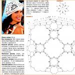 Several options for knitting a beret for beginners
 Men's dark gray noble vest
 We sew cool pillows: step-by-step master classes
 We sew a simple nightgown for beginners: everyone can do it!